
We often want to grab molecules. We may wish to extract oxygen molecules from the air we breathe, or sugars from our food, or, more deliberately, toxins from a water supply. Nature provides many pairs of molecules that bind to each other — hemoglobin and oxygen, for example — and many more have been invented. We might control whether or not the binding occurs, beyond simply controlling the presence or absence of the molecules, by tuning their concentrations and letting statistical mechanics dictate the outcome. Or we might have chemical switches, triggered for example by other molecules.
What if we want to control molecular binding with light? If the light is on, our pair binds; if off, it doesn’t. In addition to being elegant, optical control would be useful: we can control the location of light with lenses or mirrors, thereby tuning the location and timing of binding. What if we further want optical control inside living cells, for example controlling the binding of transcription factors to DNA to turn gene expression on or off? Let’s be even more demanding and ask for the tools that enable light-induced binding to be encoded in the genomes of the cells themselves, avoiding the need to deliver chemicals or tiny particles — the cell itself makes the machinery, and we just toggle the light.
This seems like an outlandish dream. It has, however, come true, and like so many developments in contemporary biotechnology and biophysics, it highlights the amazing breadth of both natural tools and human ingenuity. I started writing about this weeks ago but was spurred to finally finish by the announcement a few days ago of this year’s Nobel Prize in Physiology or Medicine, awarded for optogenetics. It’s an excellent prize choice. In brief, one controls the electrical activity of cells, such as neurons, with light. Though the goal, controlling membrane voltage, differs from the goal of molecular binding, and the molecular tools are different as well, both techniques have the obvious similarity of using light as the handle and the less obvious similarity of originating from observations of the natural world. In the optogenetics case, the creatures were light-sensitive algae. In the binding case, oats and bacteria.
Oats
Everyone with a houseplant near a window knows that plants grow towards light. As with so many phenomena, Charles Darwin was the first to document and study this “phototropism;” oat seedlings were his subject. Plants must be able to detect light and alter their cellular processes in response. How? In 1997, researchers identified in Arabidopsis thaliana, a model plant used especially for genetic studies, “LOV” (Light, Oxygen, or Voltage) domains in proteins that were known to be necessary for phototropism [1]. It was unclear what these domains were doing. Over the next five years or so various aspects of LOV signaling were figured out, most notably for our purposes the realization that the light-induced chemical change leads to a mechanical change in the protein, exposing an otherwise hidden region [2]. Even though the structural change involves a covalent bond, it’s reversible, and after some time in the dark the protein reverts to its normal shape.
This work was done in oats (Avena sativa), and oats continued to be the system of choice from which to design variants. One such variant, noted in an influential 2015 paper [3], involved cleverly embedding in the occluded region a peptide (a small chain of amino acids) called SsrA. SsRA binds strongly to another peptide, SspB. Thus we can control using light whether SsrA and SspB can bind to each other; in the dark, SsrA is hidden while in the light, SsrA is exposed and free to bind. By extention, we can control using light whether some protein “A” will bind to some other protein “B”: we link A to a LOV domain containing SsrA, and B to SspB. In the light, SsrA and SspB will bind to each other and, if there’s any affinity at all between A and B, their close proximity will favor binding. It’s as if, returning to our first paragraph, the concentration of A and B is very high; locally, via the forced connection, it is. So, if the light is on, we have a very high probability of A-B binding; if off, we don’t. (It’s even more tunable than this, in fact, since we can choose among a variety of SspB peptides with different affinities for SsrA.)
All these ingredients are simply proteins and so if we encode their genes into cells, the cells will make them. The 2015 paper [3] demonstrated that one can shine light on a small region of a cell and have A-B binding occur, as intended, there. In their demonstration, “A” was a membrane-associated protein “B” was a protein that is normally diffusing throughout the bulk cytoplasm, now locally recruited with light to the membrane. The system, by the way, was named iLID, for “improved light-induced dimer.” This is a brief glimpse of the history and I’m skipping lots of work, and skipping over the names of people who did all this work, to keep it short.
Bacteria
Where did SsrA and SspB come from? Bacteria! In 1979, a strange RNA was discovered in E. coli [4] that later became known as a transfer-messenger RNA, or tmRNA, because it has characteristics of both the transfer RNA that helps add amino acids to nascent proteins and the messenger RNAs that carry the coding sequence. Nearly 20 years later, researchers deciphered the role of this RNA: it’s recruited if protein synthesis stalls, and the tmRNA adds a specific amino acid sequence to the unfinished protein. What’s this sequence? The SsrA peptide! SsrA is recognized by SspB, another peptide made by the bacterium, and SspB also binds to a protein that destroys other proteins. Thus, if a protein is badly made, SsrA is attached, recruiting SspB and the destruction machinery; it’s a bacterial quality control system.
Thus, by paying attention to oats and bacteria — or more generally, to nature — we found ingredients to mix together for new ends, like light-induced protein binding. In an era in which funding agencies seem overly focused on science with obvious applications, it’s worth keeping in mind that most applications had their origins in basic curiosity. It’s also worth marveling at the ingenuity of the scientists who were broad enough in their interests to be aware of these ingredients. I usually don’t mix bacteria with my oatmeal.
Local activity
I learned about iLID and was reminded of LOV domains thanks to the excellent work of my University of Oregon colleagues (and down-the-hall neighbors) Scott Hansen and graduate student Sophia Doerr; I was on Sophia’s thesis committee. As described in a paper published a few months ago (“Reconstitution of Ras-PI3Kγ membrane communication and feedback using light-induced signaling inputs” [5]), the group tackled the topic of feedback in cellular signaling. In general, positive feedback can amplify responses to signals and can lead to dynamics like traveling waves. It’s thought that such feedback is important for various processes that occur at cell membranes, but it’s hard to confirm this given the complexity of actual cells. Sophia and colleagues therefore built a minimal system from the bottom up: lipid membranes, proteins, fluorescent probes, and tunable membrane recruitment via the iLID system. Adjusting the light intensity tunes the density of targeted proteins at the membrane, and patterning the light with a micromirror array patterns the protein locations.
The result: beautifully clear and quantifiable images of, for example, disks of activated protein expanding from a central, illuminated spot if the light intensity exceeds some threshold (on the right in the movie below), and fizzling out if the light is too dim or if the feedback mechanism is absent (movie, left). (If the movie doesn’t appear, try this link, or Movie 4 at the paper link above.) As always, Brownian motion is present as well, and the protein wave travels outward at a characteristic speed that is a function of the feedback-induced growth rate and the diffusion coefficient — a Fisher wave of the same sort that I wrote about in the context of wandering muskrats 10 years ago.
It’s not surprising that principles of dynamical systems arise throughout the natural world, but as a physicist, it’s always nice to see!
Another loop
We can use light to induce molecular binding and therefore spur specific biochemical or biophysical processes. One such process is the generation of light — various bacteria are bioluminescent, for example, and a commonly used protein, luciferase, consumes ATP to emit light. One can imagine, therefore, engineering a living positive feedback loop of light emission triggering light capture triggering light emission and so on… It would be horribly inefficient, but it would be an impressive feat! At a larger scale than cells, nature already shows us a living system that does this.
Today’s illustration…
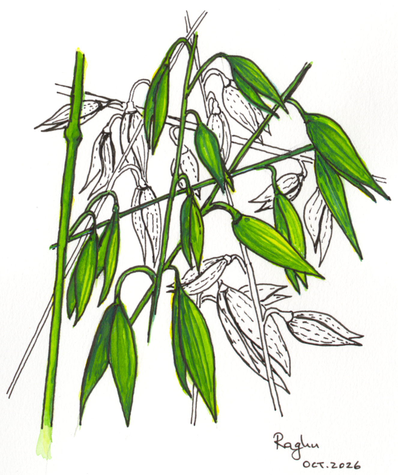
Oats!
— Raghuveer Parthasarathy, October 9, 2026
References
[1] Huala, E. et al. (1997) Arabidopsis NPH1: a protein kinase with a putative redox-sensing domain. Science 278, 2120-2123.
[2] Harper, S.M., Neil, L.C. & Gardner, K.H. (2003) Structural basis of a phototropin light switch. Science 301, 1541-1544.
[3] Guntas, G. et al. (2015) Engineering an improved light-induced dimer (iLID) for controlling the localization and activity of signaling proteins. Proceedings of the National Academy of Sciences 112, 112-117.
[4] Ray, B.K. & Apirion, D. (1979) Characterization of 10S RNA: a new stable RNA molecule from Escherichia coli. Molecular and General Genetics 174, 25-32.
[5] Doerr, S., Olavarrieta Colasurdo, A. & Hansen, S.D. Reconstitution of Ras-PI3Kγ membrane communication and feedback using light-induced signaling inputs. Nat Commun 17, 8721 (2026). https://doi.org/10.1038/s41467-026-75652-y

