

Continuing my trend of belatedly writing short descriptions of papers my group has published, this one came out in May, describing a new approach we developed for measuring the viscosity of lipid membranes:
“Measuring Lipid Membrane Viscosity Using Rotational and Translational Probe Diffusion,” Tristan T. Hormel, Sarah Q. Kurihara, M. Kathleen Brennan, Matthew C. Wozniak, and Raghuveer Parthasarathy, Phys. Rev. Lett. 112, 188101 (2014). [Link]
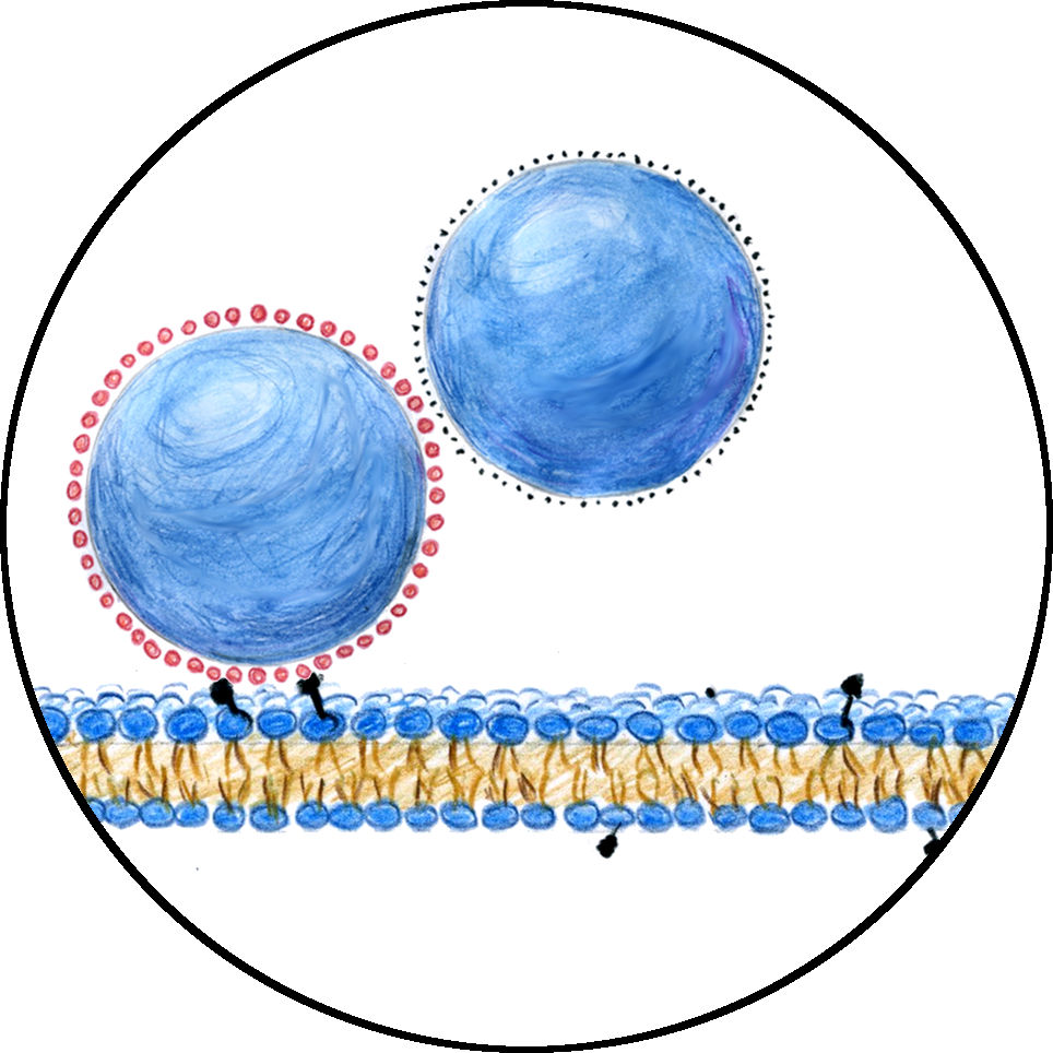
Viscosity is one of the most important material properties of any fluid, characterizing its resistance to flow (or more technically, its response to shear stresses). We’re intuitively familiar with viscosity, observing for example how warm honey flows much more easily than cold honey. For water, oils, and many other three-dimensional fluids, viscosity is well-characterized, tabulated in books and databases. For lipid bilayers, however, the two-molecule-thick liquids (illustrated above) that make up cellular membranes, viscosity is poorly quantified. It’s hard to measure, especially because lipid membranes are essentially two-dimensional fluids, whose flow behaviors differ quite dramatically from their three-dimensional counterparts. Understanding lipid viscosity is important for understanding how structures in membranes like protein clusters and cholesterol-rich “rafts” move, how proteins can alter the fluid properties of membranes, etc. In addition, it’s just embarrassing that the state of our understanding of the lipid bilayer, nature’s most important two-dimensional fluid, lags so far behind that of three-dimensional fluids.
We therefore set out to develop a new and better approach to quantifying lipid membrane viscosity. Our method hinges on Brownian motion: the random jiggling experienced by all objects due to ever-present thermal energy. As Einstein explained over a hundred years ago, the magnitude of the random motion of a particle in a liquid is a function of the liquid’s viscosity; the greater the viscosity, the lesser the motion, a relationship that holds in any number of dimensions. In fact, if one knows the size of the diffusing particle and the temperature, measuring the “diffusion coefficient” (which characterizes the random motion) is sufficient to extract the fluid’s viscosity. One can take this approach to measuring membrane viscosity, attaching particles to a membrane and watching their Brownian motion, but one runs into a problem: the effective size of the diffusing object may be different than the particle size. As illustrated below, one can imagine a variety of geometries for the particle-membrane linkage:

On the left, the effective size of the diffusing object is bigger than the particle size; on the right, it’s smaller.
We realized that in addition to the “translational” Brownian motion of particles (i.e. their meandering position), one can examine their rotational motion: the orientation of particles also shows diffusive behavior that also depends on particle size and fluid viscosity. Measuring both translational and rotational diffusion allows one to determine both the effective particle size and the viscosity. We came up with a way of making paired spherical tracer particles to link to membranes…
We can image these with fluorescence microscopy, and the pairs allow us to visualize their orientation:
Using this approach, we were able to measure lipid bilayer viscosity. Moreover, we were able to study what happens when a protein that’s involved in membrane deformation interacts with the lipid bilayer, discovering that it dramatically increases the two-dimensional viscosity — the first time such an effect has been reported.
We were very happy with how this project turned out. It was also gratifying to see that others liked it — it got chosen for a “synopsis” from the American Physical Society, one of just six for the week, and was also featured in a “research highlights” blurb in Nature Chemical Biology (?!). By a great coincidence, a paper on granular materials from my neighbor Eric Corwin also got a synopsis in the same week!
Tristan Hormel, the first author on our paper, is a graduate student in my lab, working now on a very different (and better?!) way of revealing fluid properties of lipid membranes. Sarah Kurihara, the second author, was an excellent UO undergrad biology major; she’s now doing fascinating things as a Peace Corps volunteer in Lesotho. Her blog is here: http://sarahkurihara.wordpress.com/. (I recommend it.) Katy Brennan and Matt Wozniak were great summer undergrads in the lab, here as part of a REU (Research Experiences for Undergraduates) program.
As mentioned, we’re continuing to explore the fascinating fluid dynamics of lipid membranes. We’re also using particle motions to examine viscosity in other contexts, for example inside fish guts (really), which I’ll hopefully write about in the future.

Hello, I would like to use the first picture of lipid membrane in my research paper at university if its OK with you. What do you want the reference to look like? A picture without watermark or permission to edit would be even better, I would still reference it and it wouldn’t be used online…
Miha Starič