
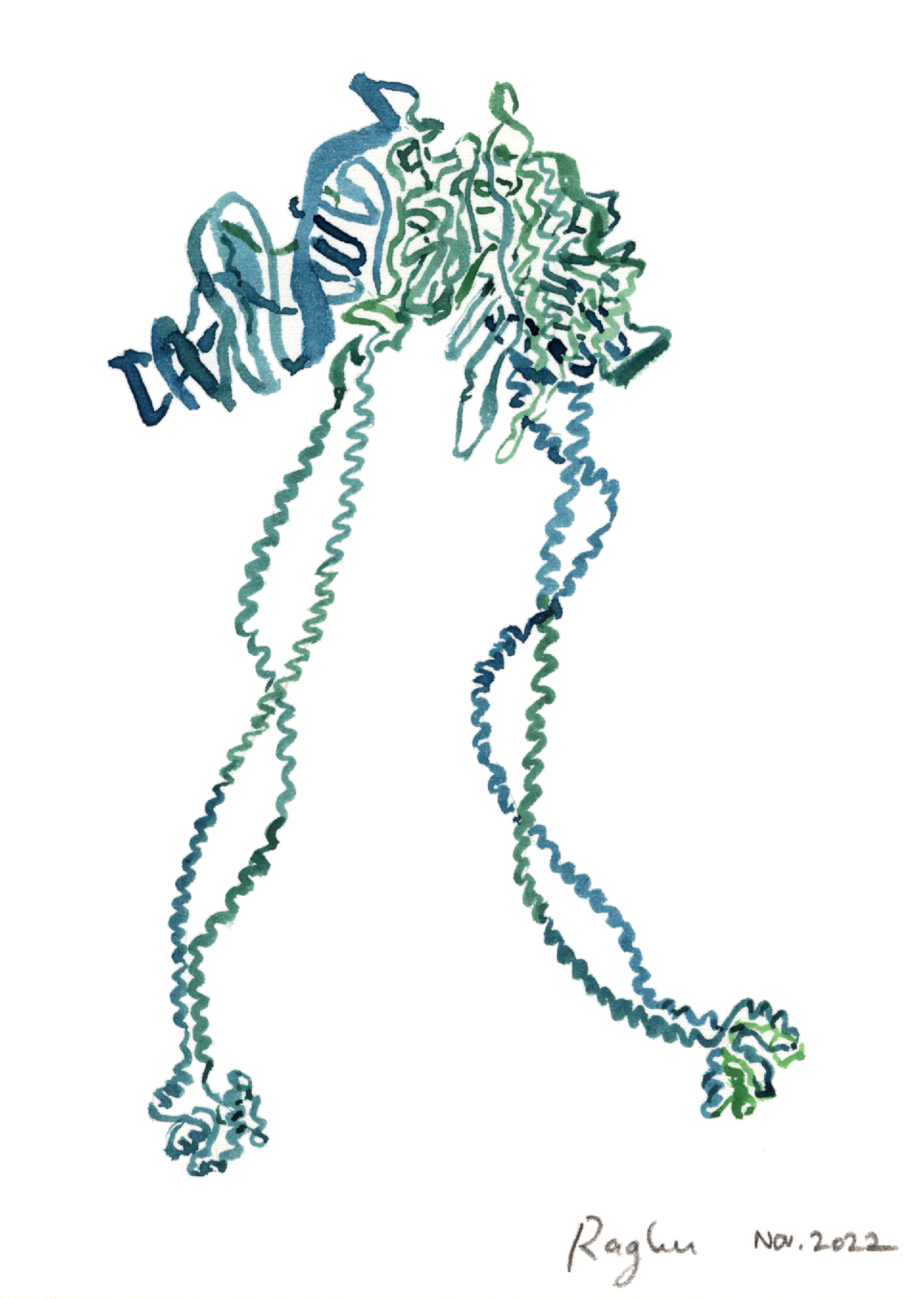
[Announcement: I’m giving I gave a virtual book talk Tuesday, Nov 29, 2022! Video recording link.] Many living things move. Panthers pounce, snakes slither, dolphins dive. Organs can move (such as beating hearts), and even individual cells can be motile, such as the immune cells that crawl through our tissues. How far down in scale we can go and still find directed, active motion — not just the passive jostling associated with temperature, but the transformation of chemical fuel into kinetic energy? The answer, it turns out, is that even molecules can propel themselves, acting as dynamic, coordinated machines. Certain proteins — the exquisitely folded chain-like molecules that form much of the substance of organisms — can perform mechanical tasks. These include spinning, forming rotary motors, and walking along tracks. “Walking” literally means walking: dynein, illustrated above, ambles along filaments made of another protein, it’s ropey legs stepping one after the other along its path. (Technically, a single dynein molecule doesn’t walk; four molecules assemble into the functional unit shown.)
There are several such motor proteins. The famous “Inner Life of the Cell” animation linked here depicts a different protein, kinesin, walking on a microtubule; this is happening right now inside all your cells. (The actual motion is much more chaotic than the animation implies.)
The ways these microscopic motors harness and transform energy, as well as the ways we can measure the associated motions and forces, are important and intensely studied biophysical topics. In addition to revealing how life works, these investigations enhance, we hope, our ability to create artificial tiny machines.
Molecular motors meander into my pop-science biophysics book, in chapters on proteins and on the ubiquity of randomness. Here are the usual links: My description, Publisher, Amazon.)
This is #12 in the series of “What is biophysics?” posts. Here’s #1 and #11.
Today’s illustration: A quick illustration of dynein, which I based mainly on Protein Data Bank structure 3VKH and slightly on the cover illustration by Graham Johnson for the March 4, 2011 issue of Science.
— Raghuveer Parthasarathy; November 11, 2022
