
 I’m a bit behind in writing summaries of recently published papers from my group. Here’s one that’s a few months old — I’m spurred to write now since I just learned two days ago that it got onto the cover of JACS, the flagship journal of the American Chemical Society:
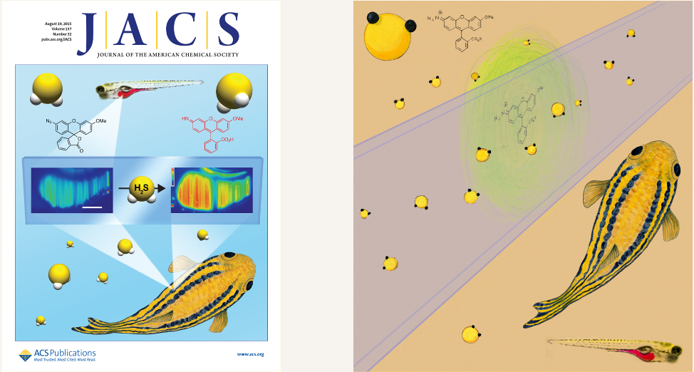
M. D. Hammers, M. J. Taormina, M. M. Cerda, L. A. Montoya, D. T. Seidenkranz, R. Parthasarathy, M. D. Pluth, “A Bright Fluorescent Probe for H2S Enables Analyte-Responsive, 3D Imaging in Live Zebrafish Using Light Sheet Fluorescence Microscopy.” J. Am. Chem. Soc. 137: 10216-10223 (2015), [link].
The cover image is the one on the left, above. (More on that in a moment.) The paper is primarily from the lab of my colleague, Mike Pluth, a remarkable organic chemist here at Oregon. Mike’s group devised a new fluorescent reporter of hydrogen sulfide (H2S) — i.e. it becomes fluorescent when it binds H2S. We’d probably all recognize H2S by its characteristic rotten egg smell. (Thankfully, my lab has never had enough of it around to notice!) There’s an increasing interest in detecting and studying hydrogen sulfide in living organisms, since it’s used by cells as a signaling molecule to regulate various physiological processes. It’s also produced by various bacterial species, and so could give insights into microbial activity.
Chemical reporters of H2S, however, have tended to be hard to use, highly toxic, or both. The Pluth lab’s new molecule is sensitive and looked like it would be amenable for use in live organisms. Since my lab does a lot of three-dimensional microscopy of larval zebrafish, we took on the task of imaging this reporter in vivo, seeing if we could detect its signal inside the larval gut. “We” is really Mike Taormina, a very skilled postdoc in my lab. This required, of course, getting the reporter molecules into the gut, which Mike did by the amazing method of microgavage — carefully inserting a fine capillary into the mouth of a larval fish, injecting the contents, and removing the capillary without damage to the fish. (A larval zebrafish is about 0.5 mm wide x a few mm long, so this procedure has to be done under a microscope.) We used light sheet fluorescence microscopy (which I’ve written about before) to image the reporter molecules, and to determine that they are properly localized in the gut. The optical sectioning capabilities of light sheet microscopy turn out to be very useful in distinguishing the gut reporter signal from the abundant background fluorescence of the zebrafish. We had hoped to detect intrinsic H2S, but the levels were insufficient for this study. Instead, we also gavaged H2S donor molecules, and detected their presence. This may seem a bit silly — detecting the very molecules we ourselves put in — but it allowed quantitative measures of sensitivity, and most importantly showed that all this could be done inside a live animal without any apparent toxicity.
In addition to showcasing the Pluth Lab’s remarkable chemical creations, the project ties into my lab’s interests in imaging not only physical processes and biological components of gut ecosystems, but also chemical activity.
After our manuscript was accepted by JACS, it was picked as an “Editor’s Choice,” and we were asked if we’d like to propose a cover image. I rather quickly painted this one as a possibility:
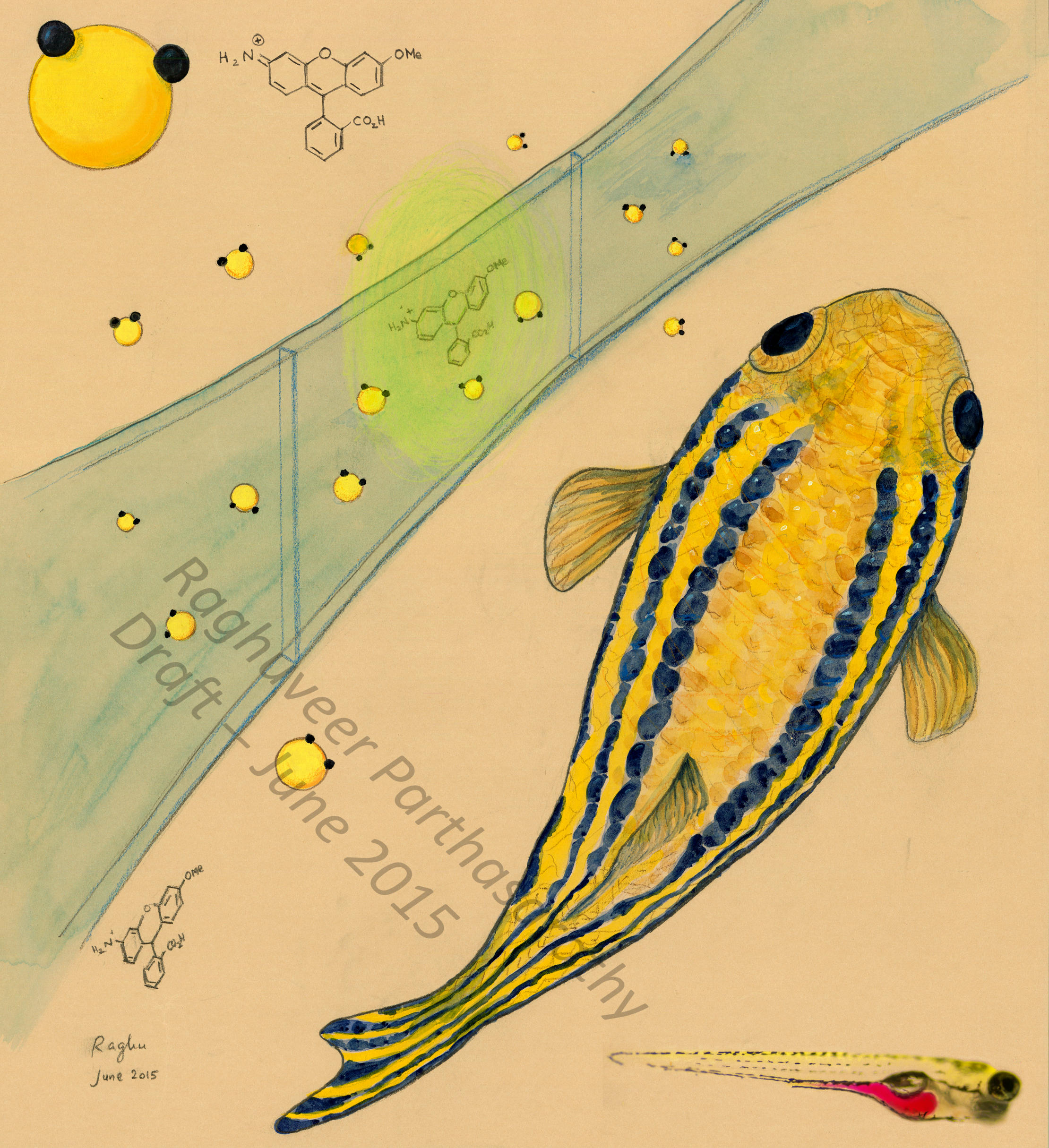 JACS suggested revising it, hence the version at the top right. It’s not great, but I rather like the fish. Usually what happens with cover art submissions is that they’re either accepted or rejected. This time, oddly, JACS was keen on having its own cover artist make a cover, which they did, but incorporating the fish from my submission as part of it. It’s a bit strange, and I have to say I’m not thrilled by the resulting cover (maybe just because I have a low tolerance for gradient shading). But still, it’s nice to have some publicity for our ability to see the smell of rotten eggs!
